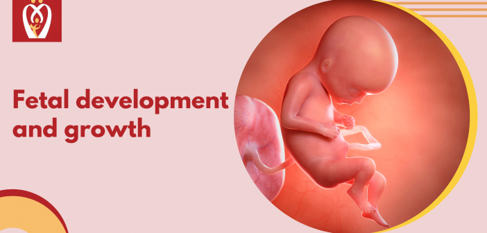
Fetal growth chart – Every expecting couple in their journey of pregnancy goes through a myriad of emotions starting with the anxiety/stress in the periconceptional period until the conception is confirmed, followed by the happiness of having a healthy fetus inside her womb on serial scans, ending with the ultimate joy of the first glimpse and cry of the healthy newborn after delivery. Through this journey, the couple has numerous questions and apprehensions about fetal development – its growth within and normality. This essay is an effort to address all these typical inquiries and allow the expectant couple to comprehend the growth of their future bundle of joy better.
Each month, a woman’s body goes through a reproductive cycle that might conclude in two ways – either a menstrual period or pregnancy. This cycle is continually going throughout the woman’s reproductive years – from puberty in her teens to menopause at the age of 50.
For a cycle to conclude in pregnancy, multiple processes/steps have to take place. First, a number of eggs (called oocytes) become ready to exit the ovary for ovulation (release of the egg) (release of the egg). The eggs grow in follicles, which are tiny, fluid-filled cysts. Imagine these follicles as little receptacles for each immature egg. One egg will develop and continue the cycle from this bunch of eggs. This follicle then suppresses the rest of the follicles in the group and the other follicles cease developing at this stage.
The mature follicle now opens and the egg is discharged from the ovary. This is termed ovulation. Ovulation normally occurs around two weeks before the next menstrual cycle starts. It typically coincides with the middle of the menstrual cycle. An embryo is formed when an egg and sperm join during the process of fertilization, which takes nine months to complete. During the menstrual cycle, the egg degenerates since there are no sperms to fertilize it.
Once the follicle is broken during ovulation, it forms a tissue known as the corpus luteum, which releases the hormones progesterone and estrogen into the bloodstream. Endometrium (uterine lining) preparation is assisted by progesterone, which aids in the onset of pregnancy. Once the egg has been fertilized, it will begin to develop in this lining. This lining is lost during the menstrual period if the lady does not get pregnant during a cycle.
Two weeks following the previous menstrual cycle, around the time of ovulation, fertilization occurs. In order to prevent additional sperm from entering, the protein covering the egg undergoes a change as the sperm penetrates. Because just one egg and one sperm are needed to create an embryo, only one may be used.
Since ovulation, the egg has been fertilized, and the real embryo or fetal age (sometimes called conceptual age) is the period of time that has transpired since that fertilization. However, since most women do not know when their ovulation has happened, but do know when their last regular menstrual cycle started, the menstrual age is utilized to calculate the age of a pregnancy. Gestational age and menstrual age are both terms used to describe the same thing. When it comes to conception, it’s roughly two weeks ahead of time. The date of conception will be a factor in determining how far along in the pregnancy the healthcare provider/obstetrician will be using this data. The gestational age is traditionally stated in weeks. Therefore, a 36-week fetus is defined as a fetus that is 36 weeks and 2 days old.
All of a baby’s genetic information is present at the time of conception, including its sex and gender. The sperm that fertilizes the egg during conception determines the baby’s gender. Women are born with the XX genetic combination, whereas males are born with the XY combination. Consequently, the mother always produces an egg with the prefix “X”. It is possible for the sperm to be an “X,” or “Y.” Two different kinds of eggs may be fertilized by different kinds of sperm, and the result is two different kinds of children: one that is female, and one that is male. (pregnancy growth chart)
The fertilized egg that will become a baby divides quickly into multiple cells, termed embryos, within 24 hours after fertilization. Embryos begin to take on the appearance of a baby during the eighth week of pregnancy and are hence referred to as fetuses. A normal pregnancy lasts 40 weeks and is separated into three trimesters. In these 40 weeks, the fetus continues to grow and develop, culminating in the baby that the expectant parents are so anxious to bring into our world.
If I am pregnant, how soon should I talk to my healthcare provider? | fetal growth chart
The majority of GPs will advise you to come in for an appointment once you have a positive home pregnancy test. Women who have enough hCG circulating in their bodies can perform these tests with great accuracy. During this stage, GPs usually prescribe a folic acid vitamin supplement, which must also be taken periconceptionally (when planning a pregnancy). In order for the baby’s neural tube (the beginning of the brain and spine) to develop correctly, the mother must get at least 400 micrograms of folic acid each day during pregnancy. Most GPs recommend taking folic acid 2-3 months before getting pregnant.
The Nurturey PinkBook is an amazing tool for all parents that gives you trusted NHS guidance about the weekly growth and development with many more features!


